INSTRUMENT: 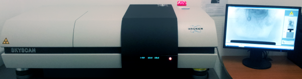
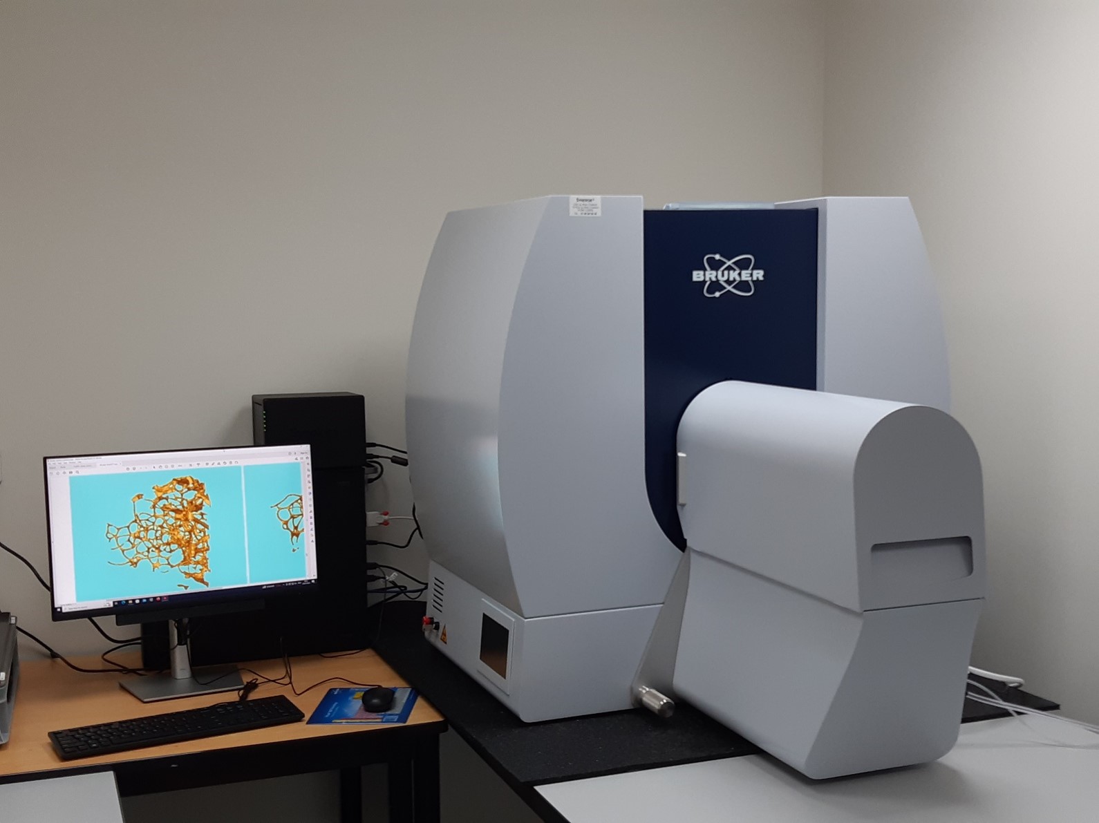
X-ray Microtomograph Skyscan 1172 100kV (Bruker mCT, Kontich, Belgium).
contact: Jérôme DELATTRE
SAMPLES:
As a non-destructive technique, X-ray microtomography is the best choice to investigate hard tissue, such as bone or teeth, in volumes at a micrometric scale.
Softer tissue might need a specific staining in order to contrast your volume of interest. In our laboratory we use osmium tetroxyde staining to show adiposity contained in the bone marrow under X-ray illumination (see images and video below).
It is possible to use Polymethyl Metacrylate resin to embed bone samples for long term conservation and easy manipulation. Softer samples are scanned with adapted tubes and holding medium.
DATA TREATMENTS & ANALYSIS:
As a tridimensionnal analysis technique, X-ray microtomography provides a quantitative analysis as measurements of volumes. Usual measurements correspond
to 2D and 3D morphometry analysis.
It’s also possible to do qualitative analysis with adapted calibration phantoms. In our laboratory we use Calcium Hydroxyapatite phantoms to do bone mineral density evaluation.
X-ray microtomography provides projections of the sample within an acquisition dataset. This dataset is used to reconstruct the volume as cross-sections. Treatment is done on cross-sections to determine a volume of interest and segmentation by binarization is used to provide the volume which will be analysed. Vizualisation might be done as MPR and volume or surface rendering.
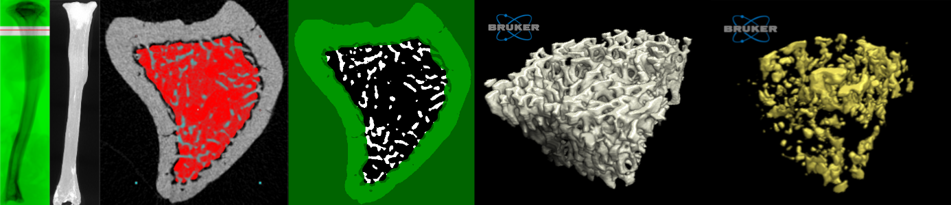
The video shows trabecular bone from rat. The trabecular bone is represented in white. The bone marrow adipose content stained with osmium tetroxyde is represented in yellow.
 INSTRUMENT:
X-ray Microtomograph Skyscan 1276 (Bruker mCT, Kontich, Belgium)
SAMPLES:
under construction
DATA TREATMENTS & ANALYSIS:
under construction